Glaucoma

Determination of glaucoma

Glaucoma is a multifactorial neurodegenerative disease of the eyes, characterized by the development and progression of glaucomone atrophy of the optic nerve with the loss of optic functions, regardless of the level of intraocular pressure (IOA) [1, 2]. An increased level of intraocular pressure can be considered as an important, but not the only pathogenetic link in the development of glaucoma.

Take of Mexidol® with glaucoma is able to reduce the
development of the development and severity of the consequences of the disease.

Types of glaucoma

There are three most frequent signs of glaucoma (Gref Triad) [3]:

  • constant or periodic increase in IOGD;
  • changes in the optic nerve;
  • Changing the fields of vision.

In origin, the primary and secondary glaucoma is distinguished.

  • Primary glaucoma is an independent eye disease. Its share is more than 80 % of all cases of glaucoma.
  • Secondary glaucoma is formed as a result of other diseases of the eye, may be the result of its injury, inflammatory diseases of the organ of vision (uveitis, keratitis), cataracts, thrombosis of the central vein of the retina, etc. Secondary glaucoma more often than primary, has unilateral localization. The clinical current is determined by the open or closed angle of the front chamber of the eye.

According to the mechanism for increasing the intraocular pressure of the glaucoma, it is divided into open -angle and closing. Primary open -angle glaucoma is a chronic progressive opticalopathy, which is characterized by the following signs [4]:

  • periodic or constant increase in the level of intraocular pressure above the individual norm;
  • structural pathological changes in the optic disk and a layer of nerve fibers of the retina; 
  • typical defects in visual fields corresponding to damage to the optic nerve disk and a layer of nerve fibers of the retina; 
  • Open angle of the front chamber (confirmed by gonioscopy) [6, 7]. 

Primary closed -angled glaucoma is a disease, in which the cause of the intraocular pressure increased the outflow of intraocular fluid due to the blockade of the angle of the front chamber with the root of the iris, accompanied by the following signs [5]: 

  • periodic or constant increase in the level of intraocular pressure;
  • optic atrophy (with excavation); 
  • Changes in the field of vision;
  • Closed angle of the front chamber.

Thus, the main difference between the open -angle glaucoma and the closure is that with an open -angle glaucoma, taking into account the open corner of the front chamber of the eye, free access of the intraocular fluid is possible, but its irregular outflow occurs, and with a closed glaucoma, the angle of the front chamber is closed, and the liquid cannot be freely circulated, which leads to an increase in intraocular pressure (Fig. 1).

Epidemiology

In the Russian Federation, about 1,250,000 patients with glaucoma aged 18 years and older, which is 1077.8 people. For 100 thousand adult population, that is, every 93 adult in the Russian Federation has glaucoma. The proportion of the first -a -examination and medical examination of diseases of the glaucoma in the adult population is more than 9 % [8]. The number of people suffering from glaucoma increases with age and is about 14 %in a group over 80 years old. In all regions of the Russian Federation, Glaucoma takes 1st place among the causes of disability due to ophthalmopathology [9]. Population studies in the world confirm the predominance of primary open -angle glaucoma with a share of 72.3–96.1 % of all forms of glaucoma and increasing its frequency with age [10]. Primary open -angle glaucoma is found more often in women (56.6 %), somewhat less often in men - 43.4 %, which correlates with the life expectancy [11]. The prevalence of primary disability due to glaucoma increased from 0.04 to 0.35 to 1000 adults, and the indicators themselves significantly vary in various territories of the Russian Federation. In most of the disabled persons by vision due to glaucoma, it was precisely the sample (80.9 %). In the contingent of disabled people, men account for 58.2 %, women - 41.8 %.

Figure 1. The mechanism for increasing intraocular pressure with open -angle
and closed -angled glaucoma

During the initial examination by disabled group I, 35.4 %were recognized, II groups of 45.8 %, group III - 18.8 %of patients with glaucoma [12]. According to some epidemiological studies, in the Russian Federation, in 40–80 % of cases, glaucoma is diagnosed in advanced stages, and in 50 % of cases, patients do not know about their disease and do not receive appropriate treatment [13].

The age of patients with first diagnosed glaucoma is mainly in the range from 35 to 90 years. The incidence rate increases in older age groups: from 40 to 49 years - 0.55–0.88 (per 1000 population), from 50 to 59 years - 1.5–2.5; from 60 to 69 years - 6.44–7.45; from 70 to 79 years - about 15.7 people; From 80 to 89 years - about 17.5 people. Thus, the number of patients with primary open -angle glaucoma with age grows exponentially, increasing in the older age group by almost 20 times [6, 7].

Etiology and pathogenesis

Currently, the primary open -angle glaucoma is considered as a complex complex of neurogramoral, hemodynamic and metabolic factors, the relationship of which is transformed over time and determines the possibility of progression of glaucomatic optical neuropathy [14, 15].

Despite numerous studies of pathogenetic mechanisms of primary open -angle glaucoma, a clear idea of the causes and progression of this disease is not formed [16]. In the explanation of its occurrence and development, the multifactorial concept prevails with the involvement of various genetic and non -generation factors [17]. As risk factors for the development and progression of primary open -angle glaucoma, they consider [4]:

  • Age: develops in patients over 40 years of age and prevalence increases with age; 
  • race/ethnicity: prevalence is several times higher in African Americans than in people of the Caucasian race; 
  • Family history: the prevalence of glaucoma among blood relatives of patients with primary open -angle glaucoma is 4–9 times higher than in the general population;
  • An increased level of intraocular pressure: directly correlates with an increase in prevalence and incidence. The risk of developing glaucoma increases by 10 % by every 1 mm Hg. Art. increasing the level of VGD above the average norm;
  • Pseudoxfoliative syndrome (chronic disease of the anterior segment of the eye from among the usepathies): the presence of pseudo -exfoliation increases the risk of glaucoma by 9–11 times. For 10 years, the glaucoma has been developing in every third patient with pseudo -exfoliative syndrome;
  • Pigment dispersion syndrome: the risk of pigmented glaucoma in patients with pigmented dispersion syndrome amounted to about 10 % in 5 years and 15 % in 15 years; 
  • The thickness of the cornea in the central optical zone (CTR): an increase in the risk of the development of primary open -angle glaucoma by 30–41 % for every 40 μm of the TSR is lower than the average norm;
  • Myopia: myopia of medium and high degree (6 diopters) increases the frequency of glaucoma;
  • Perfusion eye pressure: the relationship of low perfusion eye pressure was found with an increase in the frequency of primary open -angle glaucoma. Since perfusion pressure represents the difference between the diastolic level of blood pressure and the value of intraocular pressure, the treatment of arterial hypertension in patients with glaucoma can lead to the development of systemic hypotension, and, as a result, the deterioration of blood supply to the optic disk;
  • Hemorrhage on the optic disk. In the international glaucoma study of normal pressure (Collaboative Normal Tension Glaucoma Study, CNTGS), the presence of hemorrhages in the optic disk zone reliably correlated with the progression of glaucoma;
  • Other factors: cardiovascular pathology, including systemic arterial hypotension, systemic atherosclerosis; Vasospastic syndrome, migraine, apnea in a dream, syncopal states of unclear genesis, diabetes, etc.

The pathogenetic basis of the primary open -angle glaucoma is optical neuropathy, due to the action of various factors leading to the apoptosis of the ganglion cells of the retina [18].

Glamoil optical neuropathy

- This is the acquired pathology of the optic nerve, characterized by the progressive loss of ganglion cells of the retina and their axons and leading to structural and functional damage to the optic nerve, impaired optic functions and blindness [4]. 

Currently, there are three main theories of pathogenesis of glaucoma optical
neuropathy: mechanical, vascular and metabolic [19].

According to the mechanical theory, the start -up factor is an increase in intraocular pressure, which leads to the deflection of the lattice plate
and damage to the axons of ganglionic retinal cells. The vascular theory explains the occurrence of glaucoma optical neuropathy with a decrease in blood flow in the optic head. The main reason for the violation of its blood supply is the disorder of vascular regulation, which causes a decrease in the perfusion of the retina and impaired local self -regulation, which leads to increased sensitivity of the optic nerve to fluctuations in intraocular pressure. Mechanical and vascular factors, summing up, activate a number of metabolic processes. Ichemia of nervous tissue contributes to the formation of an excess of free radicals and the activation of the processes of lipid peroxidation processes. The cascade of pathological biochemical processes is launched, which, in turn, have a cytotoxic effect on the retina and visual nerve [19]. According to modern data from domestic
and foreign authors, there is a hypothesis that one of the main initiating mechanisms starting a chain of events leading to glaucomal damage is a violation of a universal mechanism for regulating vascular tone due to endothelial dysfunction [20]. The main manifestation of endothelial dysfunction is the imbalance of vasoconstrictor
and vasodilator endothelial mediators.

The leading among endothelial vasodilators is nitrogen oxide (No),
and among the Constructors-endothelin (ET-1). The disagreement of the action of these mediators triggers the mechanisms of adaptive distress, which leads
to the progression of morphological destruction, dyslipidemia, the development of hemodynamic and hydrodynamic disorders [21]. Azot oxide, being a powerful vasodilator, in high concentrations has a cytotoxic effect, as it stimulates the formation of free radicals
in peripheral blood. Reduced NO products causes vasospasm
in isolated ciliary arteries, which are the main source of blood supply to the optic disk, and leads to the progression of the glaucomic process [22]. The essential trigger mechanism of dysregulation, also contributing to the occurrence of endothelial dysfunction, is the development of oxidative stress in the body [23]. Oxidative stress, which is a component of neurodegeneration of subcellular structures of ganglion cells of the retina, can both show a direct cytotoxic effect
and act through signaling mechanisms, causing the death of retinal
cells [24].

Classification of primary
open -angle glaucoma [4]

The following pathogenetic forms are distinguished: primary simple glaucoma; glaucoma of low pressure; pigmented glaucoma; Pseudoxfoliative glaucoma.

VGD level


Normal (a)


Moderately increased (b)


High (c)

Tonetric ICD, pt (mm Hg)


≤ 25


26-32


≥ 33

True High School, P0
(mm Hg)


≤ 21


22-28


≥ 29

Table 1. Classification scheme of the state of intraocular pressure in glaucoma 

The course of glaucoma
Clinical characteristics
Stabilized

The absence of negative dynamics in the state of the optic nerve and field of vision
with prolonged observation of the patient (at least 6 months)

Unlocked

With repeated studies, the negative dynamics of structural (optic disk, nerve fibers of the retina) and functional indicators (a change in the field of view) are recorded

Table 2. Classification scheme of glaucoma by the nature of the course of the disease

When assessing the dynamics of the glaucomatous process, the level of intraocular pressure and its correspondence to the “target” values are also taken into account.
According to the severity of the pathological process, the following stages are distinguished (Table 3):


Stages


I, initial


II, developed


III, far away


IV, terminal

Signs

Field of view


The boundaries of the field of view are normal, small changes (scotomas) in the paracentral areas of the field of view (small paracentral cattle, relative scotoma in the Bjerrum zone)


Narrowing of the boundaries of the field of view from the nasal side by more than 10 ° or the merger of small paracentral cattle into an arcuate cattle


The boundary of the field of view from the nasal side (or concentrically) is less than 15 ° from the fixation point. This stage also includes cases with a area of vision, which has survived only on the periphery in the absence of central vision


Loss of objective vision

The optic disk


The asymmetry of excavation on two eyes, the vertical-oval form of excavation, excavation is expanded, but does not reach the edge of the optic disk


The excavation of the optic nerve disk is expanded, the regional excavation of the optic disk appears


Regional subtotal excavation of the optic disk


The marginal excavation is total

Table 3. Classification scheme of the stages of glaucoma

Clinical picture

Glaucoma, as a rule, proceeds asymptomatic, and in most cases is found at a developed and/or far -reaching stages (for example, during preventive examinations, when selecting glasses, examination of the fundus, etc.). Separate atypical complaints of patients with glaucoma, which should be paid attention to, can be: periodic clouding of vision, a vision of rainbow circles around light sources, pain in the eyes, headaches, flickering of “flies”, fatigue, as well as a frequent change in presbyopic glasses, apparent moistening of the eyes, lacrimation, etc. [25]. Tentatively, the duration of the preclinical and initial stages is from 1 to 5 years. In some cases, the disease goes through all stages up to complete blindness within 3-5 years [6]. Glaucoma is characterized by a periodic or constant increase in the level of intraocular pressure above the individual norm. The asymmetry of the ophthalmotonus between paired eyes> 2-3 mm RT is detected. Art., and the range of oscillations of the level of IOD during the day may exceed 5 mm RT. Art. [26]. The primary open -angle glaucoma is characterized by structural pathological changes in the optic disk and a layer of nerve fibers of the retina. Excavation develops and deepens on the fundus, which in the final of the disease becomes total and deep. There is a local or diffuse thinning of a layer of nerve fibers of the retina and progressive atrophy of the mesh layer [27]. Primary open -angle glaucoma is characterized by both diffuse and focal changes in the fields of vision. The initial stage of the disease is characterized by the expansion of a blind spot, focal lesions (scotomas) in the paracental department. The progression of visual defects is characterized by the presence of a new defect in a previously normal area, the deepening of a previously existing defect, the expansion of the previously existing cattle, the presence of a general decrease in sensitivity.

The final of the disease determines the residual “island” of the light -pricing on the temporal side or complete blindness [6].
The clinical picture of the primary closure glaucoma depends on the closing mechanism of the angle of the front chamber and the course of the disease [6].
Sometimes intraocular pressure is made by a sharp jump against the background of the closed corner of the front chamber of the eye. In this case, we are talking about an acute attack of glaucoma - a urgent state, capable of leading to irreversible and complete loss of vision. Since the acute attack of glaucoma can have serious consequences
for vision, it is very important to recognize the problem in a timely manner and take measures to stop it. Glaucoma is considered the provoking factors for the development of an acute attack of the gazeks, emotional stress, stay
in a darkened room, work with a head tilt, accommodation stress, hypothermia. The patient makes complaints about headache, nausea, pain
in the eye, reduction of vision, rainbow circles. During an objective examination against the background of an acute attack of glaucoma, high intraocular pressure is determined, hyperemia of conjunctiva, swelling of the cornea, swelling of the iris, small front chamber, watery moisture loses its transparency (due to protein effusion), the pupil is expanded, the pupil reaction is missing. Due to the rapid increase in ophthalmotonus and a significant compression of the rosucua root, there is a violation of blood circulation in radial vessels with signs of necrosis and aseptic inflammation. The edema of the cornea and the optic disk is noted. After an attack, focal atrophy of the iris, deformation and shift of the pupil remains. The most important sign of an acute attack of glaucoma, which can be determined with gonioscopy, is the closed angle of the front chamber of the eye. If it is open, then this is not an acute attack of glaucoma [5].

Prevention

Specific prevention of glaucoma does not exist. It is recommended to measure intraocular pressure at the first passage of a preventive medical examination, then at the age of 40 years and over 1 time per year with the aim of early detection of glaucoma [28]. An examination (consultation) by an ophthalmologist of citizens aged 40 and older, having increased intraocular pressure, and citizens aged 65 years and older, have a decrease in point of vision, not amenable to point correction, with the aim of early detection of primary open-angle glaucoma [1, 24].
With a stabilized course of glaucoma, it is necessary to carry out visimometry, ophthalmotonometry, ophthalmoscopy, perimetry at least once every 6 months, gonioscopy - at least once a year. Patients with an unstabilized course of the disease need individual surveillance periods - depending on the characteristics of the course of the glaucy process, the presence of concomitant pathology and the drugs used [6, 28]. If there is a technical possibility, it is advisable to conduct optical coherent tomography once every 6-12 months.
At each visit of the ophthalmologist, an assessment of the patient’s subjective well -being, his visual functions (difficulties in driving, dark adaptation, problems with contrast sensitivity, reading a small font and assessing the distance to objects), the quality of life and commitment of the patient with prescribed treatment are carried out. Ophthalmotonometry is carried out by the same method at each examination of the patient (taking into account the time of day) with an assessment of the presence or absence of the level of pressure pressure.

In the absence of a target of target or fluctuation of indicators, ophthalmotonometry may be required at different times of the day. Honioscopy is carried out by the patients with primary open -angle glaucoma at least once a year, more often at the risk of closing the angle of the front chamber. In case of perimetry, changes in the field of view should be confirmed by at least one repeated test [4].
For patients with glaucoma when going to a doctor for any disease, it is necessary to inform a specialist about observing an ophthalmologist in connection with a glaucoma, since there are a number of drugs contraindicated in this disease. These include some antidepressants, anti -parquinsonic drugs, vasoconstrictor eye drops, anticonvulsants, some analgesics, etc. In any forms of glaucoma, the use of table salt, pickled and fatty dishes is contraindicated. The regular use of alcohol is prohibited. All activities associated with tilts down, for example, work in the country, are prohibited. Some types of fitness and sports are also not recommended. Contraindicated exercises associated with lifting weights and jumps. A long stay in the dark negatively affects the hydrodynamics of the eye, so it is necessary to read, watch TV, work and perform daily household matters with a fairly bright light. A long stay in the dark or twilight is contraindicated. In the summer, when entering the street, it is recommended to use sunglasses with high-quality UV filters. The long -term stay of patients with glaucoma in the cold is not allowed, especially in the wind, as well as under direct sunlight.

Treatment

All patients with primary open -angle glaucoma are recommended to reduce
the level of intraocular pressure in order to prevent the progression
of glaucoma of optical neuropathy [6, 29].

The level of intraocular pressure can be reduced using local drug therapy, laser treatment or surgical methods (possible combination of methods). Surgical treatment is used when it is impossible to reduce intraocular pressure with the help of drug methods, and changes in the optic nerve disk and the state of ganglion cells of the retina are progressed [30]. Prolonged use of drugs leads to a decrease in the antihypertensive effect, which requires timely correction of the therapy when identifying signs of the subcompensation of ophthalmotonus [31]. Prior to the appointment of treatment, the alleged level of “target” pressure should be determined taking into account the initial values of ophthalmotonus, the stage of the disease, the potential rate of its progression, age and expected life expectancy, as well as additional risk factors. All patients with primary open -angle glaucoma need to strive for the maximum decrease in the level of intraocular pressure based on the stage of the disease.

An individual approach to the treatment of glaucoma is to adapt to the needs of a particular patient. Patients with a pronounced decrease in visual functions or young patients with the manifestation of the disease should receive more aggressive treatment and are more closely observed than patients with a low risk of deterioration of visual functions [32]. Of the drug therapy, preference is given to local drugs, as a rule, in the form of eye drops. Local therapy should be aimed at reducing intraocular pressure. As preparations of the first choice, analogues of prostaglandins and prostamids, which have maximum hypotensive activity, are used. Preparations of other pharmacological groups (local carboanhydrase inhibitors, alpha-2-adrenosimetics, selective beta-blockers) are used less often as their lower hypotensive efficiency [4].

In the mechanism of the death of ganglion cells of the retina, many
factors and biochemical reactions are involved (see the section “Etiology and pathogenesis”). A decrease
in intraocular pressure does not always help stop
the progression of the glaucomal process. Thus, it is necessary to use
drugs that allow you to protect and prevent damage to the layer of nerve
fibers of the retina [30].

Such drugs include drugs with antihyplance and antioxidant action mechanism - neuroprotectors [6]. Neuroprotection in glaucoma involves the correction of general and local hematocycrians and neurodistrofic disorders, i.e., it provides the maximum possible protection of the retina and optic nerve from the damaging effects of mechanical and vascular factors. The use of neuroprotectors allows you to stabilize the visual function and thereby slow down the process of neurodegeneration [19]. In addition, in the treatment of patients with primary open -angle glaucoma, drugs with a multimodal (multidirectional) mechanism of action are necessary, including wide potential for correcting endothelial dysfunction [33]. According to national clinical recommendations, the condition for prescribing is the presence in the instructions of the drug The indication “primary open -angle glaucoma”, which guarantees its safety and effectiveness in this disease [4]. A bright representative of drugs with a multimodal action mechanism is Mexidol® (original ethylmethylhydroxypyridine succinate). Mexidol® is a unique development of the domestic pharmaceutical industry and has a 25-year clinical experience in successful use in various fields of medicine [34]. The mechanism of action of Mexidol is due to a combination of pharmacological effects: antioxidant, antihyplance and membrane -resistant. Penetrating through the hematoencephalic barrier, Mexidol® enters the nerve cells and concentrates in mitochondria, which ensures its high pharmacological activity. Mexidol® helps to preserve ganglion cells of the retina and optic fibers with progressive neuropathy, the causes of which are chronic ischemia and hypoxia, improves the functional activity of the retina and optic nerve [35]. The instructions for the medical use of the drug Mexidol® as one of the indications for use indicate the primary open -angle glaucoma of various stages as part of complex therapy [36]. 

A high degree of effectiveness and a favorable safety profile of the drug Mexidol® in patients with primary open -angle glaucoma are confirmed by the results of clinical studies [16, 35, 37]. In order to study the effectiveness and safety of the drug Mexidol® as part of the complex therapy of primary open -angle glaucoma under the leadership of the village of M., Professor E. A. Egorov (2011), an open, randomized controlled comparative study was attended, in which 94 patients aged 18 to 75 years with primary open -angle glaucoma of the I - III stages took part. Patients were divided into 3 groups: 50 patients were prescribed combined treatment of 100 mex of Mexidol intramuscularly and 150 mg of picclone, 22 patients took 300 mg of Mexidol intramuscularly and 150 mg of picclone, 22 patients received only 150 mg of picclon. Patients took Mexidol® once a day, the duration of the intake depended on the stage of glaucoma: 14 daily therapy - for the 1st and 2nd stage of glaucoma, 21 for the 3rd stage of glaucoma. The examination included standard ophthalmological, as well as perimetry, electroretinography, the study of the arterial blood flow of the retina and the optic disk. According to the results of the study, Mexidol® showed antihypoxic, antioxidant, nootropic and membraneoprotective effects, which is reflected in the improvement of the studied indicators and visual functions. Mexidol® has a positive effect on the course of primary open -angle glaucoma not only in combination with other systemic drugs, but also in the form of monotherapy. To achieve the best result of treatment for patients with primary open -angle glaucoma of the I - III stages, Mexidol® should be prescribed at a dose of 300 mg/day. intramuscularly or intravenously dropped in 400 ml of sodium chloride solution daily for 14 days. It is possible to combine the drug Mexidol® with drugs that have nootropic effects [37].


In the study of the village of M. E. S. Leonova (2015) assessed the effectiveness of the consistent prescription of the drug Mexidol® to patients with primary open -angle glaucoma of the I - III stage with a compensated level of intraocular pressure. Mexidol® was prescribed 250 mg intravenously drip for 5 days in the daily hospital mode, then an on -anchor 125 mg 3 times a day for 3 months. During the study, it was found that the long -term use of Mexidol® is effective for neuroprotection in patients with the I - II stage of primary open -angle glaucoma and suggests stabilization of the process in patients with the III stage of the disease. The inclusion in the comprehensive conservative treatment of patients with glaucoma tablet form of Mexidol allows the maximum to realize the membrane, nootropic, antihipoxic and antioxidant effects of the drug, which prevents the progression of the pathological process. Mexidol® in a tablet form is well tolerated by patients and practically does not cause undesirable reactions when taking [35]. 


The purpose of the study of the village of M. N. T. N. Malishevskaya (2023) was the study of vasomic, antioxidant and antihypoxic effects of Mexidol® in patients with primary open -angle glaucoma. The study was attended by 78 patients with the primary open -angle glaucoma of the initial stage (n = 43) and the developed stage (n = 35), whose average age was 67.8 ± 1.5 years; 47 patients of the main group in addition to local hypotensive treatment received the drug Mexidol® at a dose of 300 mg/day. Intramuscularly 1 time per day for 14 days, 31 patients compiled a control group. During the study, Mexidol® proved himself as an effective endothelioprotector. The drug had a positive vasomic effect on vascular endothelium in patients with primary open -angle glaucoma, increasing the ability of the vascular wall to vasodilatation and normalizing the ratio of bio -effective mediators; contributed to the stabilization of neuronal activity of the retina, reducing ischemic processes and thereby improving interneuronal conduct. Mexidol® significantly reduced the severity of oxidative stress, reduced the plasma level of lipid peroxidation products, and also increased the antioxidant activity of blood. In patients receiving Mexidol®, stabilization and prolonged preservation of visual functions was noted [16].

How to take Mexidol® with glaucoma?

It is recommended to take Mexidol with primary open -angle glaucoma according to the following scheme: Mexidol® intravenously drip (200-300 mg) once a day for 14 days (phase of saturation with the therapeutic effect), then Mexidol® Fort 250 in a dose of 250 mg (1 tablet) 3 times a day for 60 days (phase of maximization of the therapeutic effect).
In order to strengthen and retain the clinical effect, courses are recommended according to the above scheme 2-3 times a year.

Block of articles on this topic

The value of mitochondrial dysfunction in stabilization

Authors:
Vlasova A.S. 1.4* , Malishevskaya T.N. 2* , Petrov S.A. 1.3* , Gubin D.G. 5* , Petrov S.Yu. 2* , Filippova Yu.E. 1*

1* ANCOO DPO “West Siberian Institute of Postgraduate Medical Education”, Tyumen, Russia;
2* FSBI "NMIC of eye diseases named after Helmholtz »Ministry of Health of Russia, Moscow, Russia;
3* FGBUN "Federal Research Center of the Tyumen Scientific Center of the Siberian Branch of the Russian Academy of Sciences", Tyumen, Russia;
4* Gauz of the Tyumen region "Regional ophthalmology dispensary" of the Ministry of Health of Russia, Tyumen, Russia;
5* FSBEI in "Tyumen State Medical University" of the Ministry of Health of Russia, Tyumen, Russia

The effect of antioxidant therapy on some pathogenetic factors of primary open -angle glaucoma

Authors:
T.N. Malishevskaya 1 , Yu.E. Filippova 2

1 FSBI "National Medical Research Center of Eye Diseases named after Helmholtz »Ministry of Health of Russia, Moscow, Russia;
2 GAUZ “Regional Ophthalmological Dispensary”, Tyumen, Russia

The possibilities and results of the use of antioxidant therapy in ophthalmological practice

Authors:
A.B. Movsisyan 1.2 , J.G. Oganezova 2.3 , E.A. Egorov 2

1 GBUZ “Hospital for Wars of Wars No. 2 DZM”, Moscow, Russia;
2 FGAOU in Russian National Research Medical University named after N.I. Pirogov »Ministry of Health of Russia, Moscow, Russia;
3 FGBNU "Medical and Genetical Scientific Center named after Acad. N.P. Bochkova ", Moscow, Russia

The use of Mexidol in the treatment of primary open -angle glaucoma

Authors:
I.A. Loskutov, O.M. Andryukhina, A.A. Kovrizhkina

Moscow Regional Research Clinical Institute. M.F. Vladimir

The experience of neuroprotective therapy of primary open -angle glaucoma based on the use of various forms of Mexidol

Authors:
E.S. Leonova 1.2 , S.V. Polyakov 1.2 , M.A. Pozdnyakova 2 , E.P. Yarygina 3 , S.O. Semisinov 2

1 NUZ "Road Clinical Hospital at the Gorky station OJSC" Russian Railways ", the interdorrutal center of ophthalmology
2 GBOU VPO" Nizhny Novgorod State Medical Academy "
3 GBUZ but" City Hospital No. 35 ", City Glacks Center

List of literature

1. Basic and Clinical Science Course. Section Glaucoma / Ed. Ca Girkin. San Francisco: AAO, 2018: 262.
2. Shaarawy TM, Sherwood MB, Hitchings Ra, Crowston JG Glaucoma: Medical Di-Agnosis and Therapy (VOL.1). London: Elsevier, 2015: 674.
3. Movsisyan A. B., Kuroyedov A.V. Diagnostics of glaucoma at the present stage. Clinical ophthalmology. 2023; 23 (1): 47-53. DOI: 10.32364/2311-7729-2023-23-1-47-53.
4. Clinical recommendations “Primary open -angular glaucoma”, 2024
5. Clinical recommendations “Primary Closing Clothing Glaucoma”, 2024
6. National Glaucom Guide for practitioners. Ed. 4th, bro. and add. /Ed. E. A. Egorova, V.P. Erichev. M.: GEOTAR-Media, 2019: 384.
7. Glaucoma: Diagnosis and Management. Methods, Evidence and Recomminess. NICE: NICE, 2017: 324.
8. Statistical materials of the Ministry of Health of the Russian Federation of the Department of Analysis, Forecast, Development of Health and Medical Science of the Federal State Budgetary Institution “Central Research Institute of Organization and Information of Health” of the Ministry of Health
of the Russian Federation, 2021
. O., Gusarevich O. G., Shcherbakova L.V., Mazdorova E.V., Malyutin S.K. The prevalence of ophthalmological diseases in a population sample older than 50 years. Bulletin of ophthalmology. 2020; 136 (3): 106–115.
11. Tham YC, Li X., Wong Ty et al. Global Prevalence of Glaucoma and Projections of Glaucomaburden Through 2040: A Systematic Review and Meta-Angysis. OPHTHALMOLOGY. 2014; 121 (11): 2081–2090.
12. Neroev V.V., Mikhailova L. A. ophthalmological incidence in Russia. / in the book. "Ophthalmology. National leadership "under. Ed. S. E. Avetisova, E. A. Egorova, L.K. Moshetova, H.P. Takhchidi. M.: Geotar-Media, 2018: 15–19.
13. Neroev V.V., Kiseleva O. A., Immortal A. M. The main results of the multicenter study of the epidemiological characteristics of the primary open -angle glaucoma in the Russian Federation. Russian ophthalmological journal. 2013; 6 (3): 4–7.
14. Malishevskaya T.N., Astakhov S. Yu. The reactivity of vascular endothelium in elderly patients with primary open -angle glaucoma and physiologically aging people, depending on the severity of endothelial dysfunction. Regional blood circulation and microcirculation. 2016; 15 (4): 59–67.
15. Zagidullina A. Sh., Nugmanova A. R. The influence of disorders of systemic hemodynamics on the development of open -angle glaucoma. Medical Bulletin of Bashkortostan. 2018; 13 (1): 116–121.
16. Malishevskaya T.N., Filippova Yu. E. The influence of antioxidant therapy on some pathogenetic factors of primary open -angle glaucoma. Bulletin of ophthalmology. 2023; 139 (4): 35–43.
17. Abu-amero K., Kondkar AA, Chalam KV An updated Review on the Genetics of Primary Open Angle Glaucoma. Int. J. Mol. SCI. 2015; 16 (12): 28886–28911.
18. Kurysheva N.I. Glaucomic optical neuropathy. M: Medpress-Inform 2006; 136 p.
19. Egorov E.A., Gvetadze A.A., Davydova N. G. Antioxidant drug in neuroprotective therapy for glaucoma // Bulletin of ophthalmology, No. 2, 2013, pp. 67–69.
20. Kurysheva N.I., Irthova E. Yu., Yasamanova A.N., Kiseleva T. N. Endothelial dysfunction and platelet hemostasis with primary open -angle glaucoma. National Journal of Glaucoma. 2015; 14 (1): 27–36.
21. Filippova Yu. E., Malishevskaya T.N., Komiychuk S. N., Gubin D. G., Vlasova A. S. The severity of endothelial dysfunction, oxidative stress, lipid metabolic disorders, a decrease in the elastolastic properties and tone of peripheral vessels in patients with different options for the course of primary open -angled glaucoma depending on, depending on Polymorphism of the genes of biological watches. Russian ophthalmological journal. 2022; 15 (1): 78–88.
22. Gumanova N. G. Oxid Azot, his circulating NOX metabolites and their role in the functioning of the human body and the prediction of the risk of cardiovascular death (part I). Preventive medicine. 2021; 24 (9): 102–109.
23. Barja G. The Mitochondrial Free Radical Theory of Aging. Prog Mol Biol Transl Sci. 2014; 127: 1–27.
24. Malishevskaya T.N., Kiseleva T. N., Filippova Yu. E., Zaitsev M. S., Vlasova A.S., Nemtsova I.V., Lugovkina K. V. The state of antioxidant status and lipid blood spectrum in patients with different options for the course of primary open -angled glaucoma. Ophthalmology. 2020; 17 (4): 761–770.
25. Chen Mf, Kim Ch, Coleman Al Cyclodestrective Procedures for Refractory Glaucoma. Cochrane Database Syst Rev. 2019; 3: CD012223.; Tóth M., Shah A., Hu K., Bunce C., Gazzard G. Endoscopic Cyclophotocoagulation (ECP) For Open Angle Glaucoma and Primary Angle Closure. Cochrane Database Syst Rev. 2019; 2: CD012741.
26. Russian Glack School. Conference "Glaucoma: Theory and Practice" Collection of scientific papers:/ Edited: Acad. Ramn prof. V.N. Alekseeva, Assoc. V.I. Sadkova-St. Petersburg: Publishing House "Man and His Health", 2013.-138 p.
27. Khaw Pt, Chiang M., Shah P. et al. Enhanced Trabeculectomy: The Moorfields Safer Surgery System. DEV OPHTHALMOL. 2017; 59: 15–35.
28. Order of the Ministry of Health of Russia dated 13.03.2019 No. 124n “On approval of the procedure for a preventive medical examination and medical examination of certain groups of the adult population”.
29. The Agis Investigators. The Advanced Glaucoma International Study (Agis), 7: The Relationship Between Control of IntraoCular Pressure and Visual Field Deterior. Am J Ophthalmol 2000; 130 (4): 429–440.
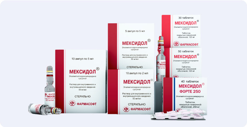
30. Loskutov I.A., Andryukhina O. M., Kovrizhkina A. A. The application of Mexidol in therapy of primary open -angle glaucoma. Effective pharmacotherapy. No. 1, 2022; 18, pp. 60–64.
31. Lichter PR, Musch DC, Gillespie BW et al. Interim Clinical Outcomes in the Collaboative Initial Glaucoma Treatment Study Comprehend Inithal Randomized to Medicates Or Surgery. OPHTHALMOLOGY. 2001; 108 (11): 1943–1953.
32. European Glaucoma Society Terminology and Guidelines for Glaucoma (4th Edition). Savona: PublicMMM, 2014: 196
. Bulletin of ophthalmology. 2019; 135 (2): 83–92.
34. Voronina T. A. Mexidol: a spectrum of pharmacological effects. Journal of neurology and psychiatry named after S. S. Korsakova. 2012; 112 (12): 86–90. Voronina Ta Mexidol: Spectrum of Pharmacological Effects. Journal of Neurology and Psychiatry Named after SS Korsakov. 2012; 112 (12): 86–90. (In Russian.).
35. Leonova E. S., Polyakova S.V., Pozdnyakova M.A. et al. The experience of neuroprotective therapy of primary open -angle glaucoma based on the use of various forms of Mexidol. Bulletin of ophthalmology. 2015; 131 (6): 91–94.
36. Https://lk.regmed.ru/register/eaeu_smpc
37. Egorov E.A., Davydova N. G., Romanenko I. A., Novikova N. D. Mexidol in the complex treatment of glaucoma // Clinical ophthalmology, volume 12, No. 3, 2011, p. 3-6.

THE INFORMATION IS INTENDED FOR HEALTHCARE AND PHARMACEUTICAL PROFESSIONALS. THIS INFORMATION IS NOT INTENDED AS A SUBSTITUTE FOR MEDICAL ADVICE.

Source of photos and images Shutterstock.com